Office Address
#07-27 Midview City 18 Sin Ming Lane Singapore 573960
Phone Number
+65 68022513
Email Address
support@everestlinks.com

#07-27 Midview City 18 Sin Ming Lane Singapore 573960
+65 68022513
support@everestlinks.com
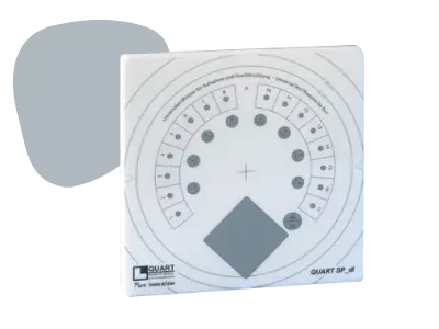
ENHANCED PHANTOM FORMAT: 330 x 330 cm
DESIGN APPROACH: Simplified design for precise artefact detection
DYNAMIC STEP WEDGE: 7 Steps; thickness 0 – 2.3 mm
SIGNAL NORMALISATION: Homogeneous area in the phantom center, 10 x 10 cm
HIGH-CONTRAST RESOLUTION: Line Pair Bar Pattern (Type 38 / Pb 0.05 mm / 45°)
LOW-CONTRAST RESOLUTION: 6 Test objects (Aluminium discs; 0.1 – 0.7 mm)
X-RAY FIELD ALIGNMENT: Field size markings for all major fields-of-view
CENTER: cross-marker, also visible when QUART ZTB beam alignment tool is in use
VERTICAL POSITIONING: Wire mount system available for tests of wall-mounted units
SIZE: 330 x 330 x 10 mm (L x W x H)
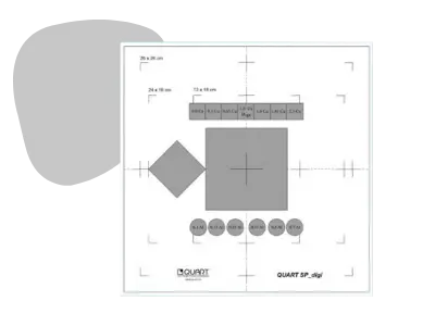
Internal Filtration: 1.5 mm standard compliant copper filtration, 17 mm PMMA as tissue simulation
Dynamic Step Wedge: 17 Steps; thickness 0 – 3.5 mm
Low-Contrast Resolution: 8 Test objects (Aluminium; 0.4 – 4 mm; Ø 15 mm), 17 Additional test objects; 1 object per step (Ø 4 mm)
High-Contrast Resolution: Line pair bar pattern (Type 38 / Pb 0.05 mm / 45°)
kV Stability: Unique kV test object (Yb + Pb)
X-RAY Field Alignment: Field size markings
Center: Radio-opaque center marker
Signal Normalisation: Homogeneous area in phantom center
Vertical Positioning: Wire mount system available for tests of wall-mounted units
Size: 200 x 200 x 18.5 mm (L x W x H)
Large Fields: Extension available to provide a homogeneous surface and field markings for formats up to 33 cm x 33 cm